
Glaucoma
What Is Glaucoma?
Glaucoma is an eye condition that damages the optic nerve. Glaucoma occurs when fluid builds up in the eye, putting pressure on the optic nerve. This pressure can damage the nerve fibers and cause vision loss.
The two main types of glaucoma are open-angle glaucoma and angle-closure glaucoma.
Open-angle glaucoma is the most common type of glaucoma. It is caused by a build-up of pressure in the eye. This pressure can damage the optic nerve over time. Open-angle glaucoma usually develops slowly and may not cause any symptoms early on. However, as the disease progresses, you may start to have problems seeing clearly, especially at night.
Angle-closure glaucoma is a less common type of the disease. It is caused by a blockage in the drainage canals in the eye. This build-up of pressure can damage the optic nerve quickly and lead to blindness. Angle-closure glaucoma often causes sudden symptoms, such as eye pain, redness, blurred vision and halos around lights.
During the early phases of glaucoma, many patients do not experience any symptoms. As glaucoma progresses, patients may experience a loss of peripheral or side vision, as well as acute eye pain, headache, blurred vision, or halos around lights.

Glaucoma
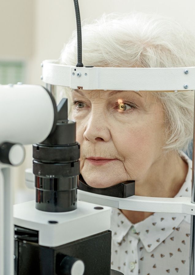
Since patients with glaucoma do not experience symptoms until later stages of the disease, a diagnosis may only be made during a standard eye checkup. Patients, particularly those with a higher risk of developing glaucoma, should have their eyes examined at least once a year to ensure that they do not develop the disease.
We may perform multiple eye tests to discover indicators of glaucoma. Visual field testing is one of the most frequent tests, and it involves drawing a map to assess peripheral vision. Blind spots in the peripheral vision are frequently indications of eye diseases, such as glaucoma.
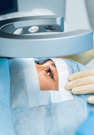
The Cirrus Optical Coherence Tomography (OCT) is a leading diagnostic technology. At Envision Eye Institute, we use this testing method to capture highly detailed pictures of the eye’s structures and tissue to assess retinal thickness and health, macular density, optic nerve integrity, and more without physically having to touch the eyes.
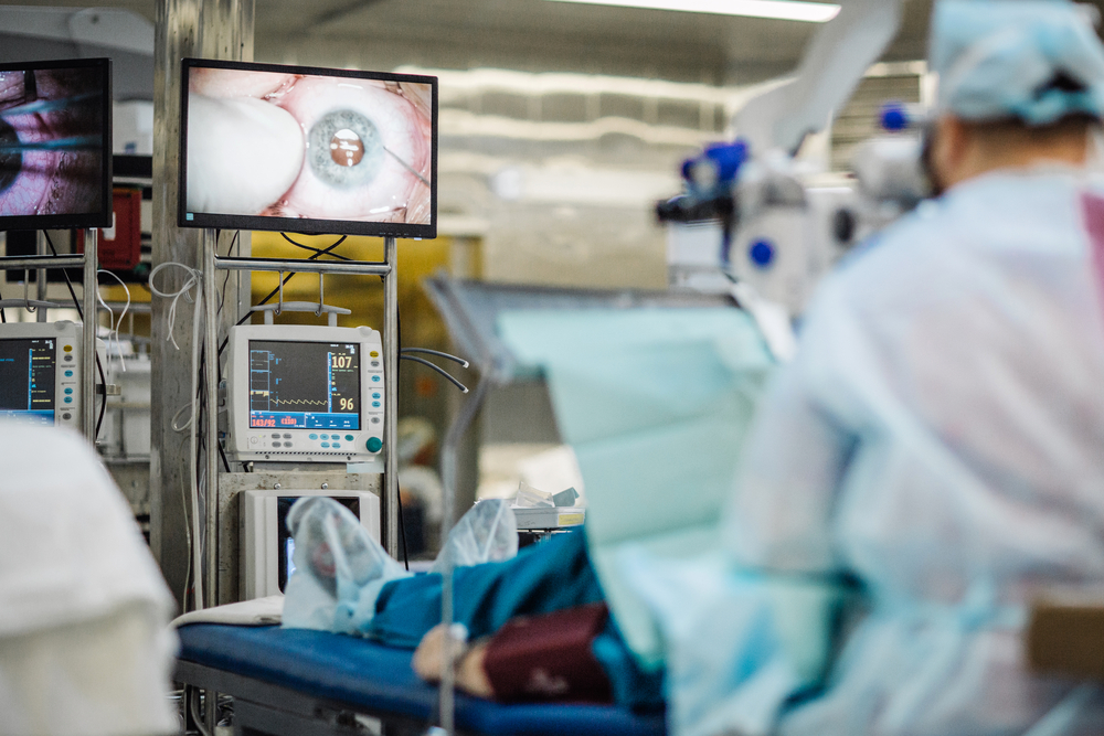
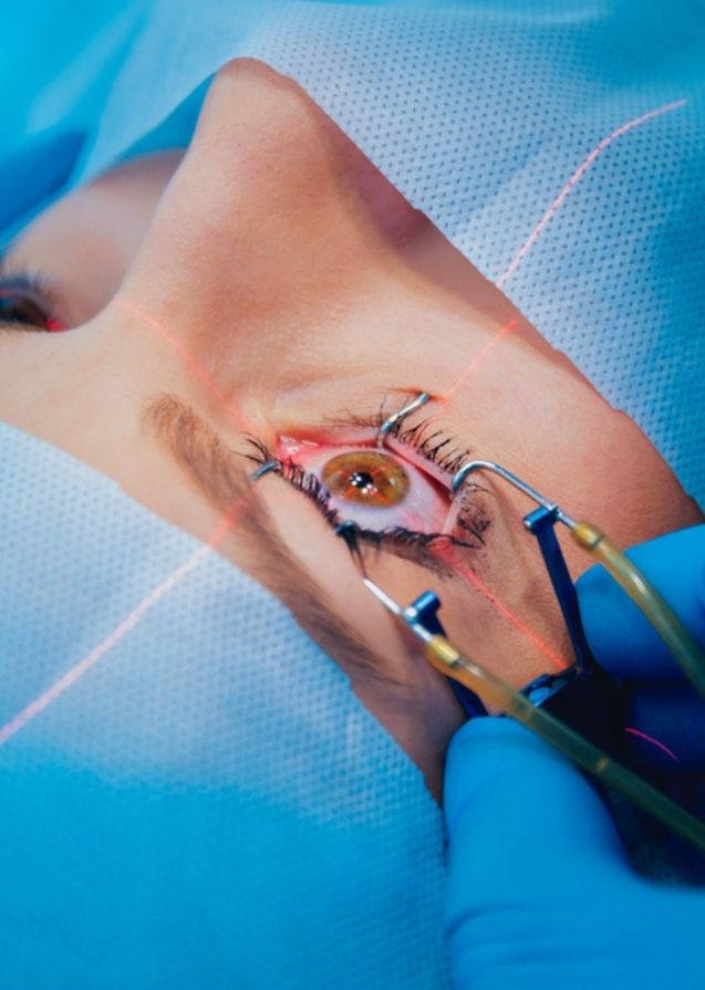
Some common treatment methods include eye drops, surgery and laser therapy such as Laser peripheral iridotomy (LPI) and Selective laser trabeculoplasty (SLT).
LPI works by creating a small hole in the iris (the colored part of the eye). This hole allows fluid to flow more freely through the drainage angle and helps to lower eye pressure. SLT works by shrinking the muscles around the drainage angle and helping to improve the flow of fluid out of the eye.
Why Us?

Latest Technology
Industry-leading vision correction treatments and procedures

Patient-centered Care
Advanced and personalized eye care for you and your family
Board-certified Surgeons
Leading ophthalmologists with extensive experience

Book Your Appointment Today
Let us improve your quality of life through proper vision care.
Contact Us